Category:Human clavicle
Jump to navigation
Jump to search
Subcategories
This category has the following 10 subcategories, out of 10 total.
3
- 3D data of human clavicle (3 F)
A
- Animations of human clavicle (14 F)
F
H
- Human sternoclavicular joint (11 F)
S
T
- Tube tops (1 P, 63 F)
V
- Videos of human clavicle (2 F)
Media in category "Human clavicle"
The following 133 files are in this category, out of 133 total.
-
A treatise on dislocations and on fractures of the joints (1824) (14753370116).jpg 2,418 × 3,050; 716 KB
-
American quarterly of roentgenology (1909) (14570815688).jpg 2,062 × 2,072; 1.44 MB
-
AP radiograph demonstrating companion shadow of the clavicle.jpg 2,016 × 2,106; 261 KB
-
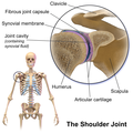
Blausen 0797 ShoulderJoint.png 2,000 × 2,000; 11.45 MB
-
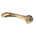
BodyParts3D Clavicle.stl 5,120 × 2,880; 113 KB
-
Chest and Neck.png 7,000 × 5,000; 31.05 MB
-
Chest skin and light.png 4,924 × 7,378; 54.28 MB
-
Chronische ventrocraniale Schulterluxation 87jm - Roe 2Eb - 001.jpg 2,217 × 1,692; 264 KB
-
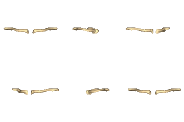
Clavicle - animation.gif 450 × 450; 1.63 MB
-
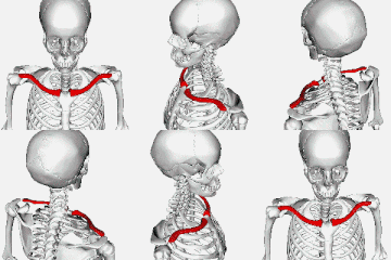
Clavicle - animation2.gif 450 × 450; 1.76 MB
-
Clavicle - animation3.gif 450 × 450; 1.87 MB
-
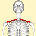
Clavicle - anterior view.png 4,500 × 4,500; 2.75 MB
-
Clavicle - anterior view2.png 4,500 × 4,500; 2.43 MB
-
Clavicle - anterior view3.png 4,500 × 4,500; 2.58 MB
-
Clavicle - close-up - animation.gif 450 × 450; 251 KB
-
Clavicle - close-up - anteiror view.png 4,500 × 4,500; 387 KB
-
Clavicle - close-up - infeiror view.png 4,500 × 4,500; 1.01 MB
-
Clavicle - close-up - inferior view animation.gif 450 × 450; 405 KB
-
Clavicle - close-up - posteiror view.png 4,500 × 4,500; 414 KB
-
Clavicle - close-up - supeiror view.png 4,500 × 4,500; 1.05 MB
-
Clavicle - close-up - superior view animation.gif 450 × 450; 315 KB
-
Clavicle - lateral view.png 4,500 × 4,500; 1.93 MB
-
Clavicle - lateral view2.png 4,500 × 4,500; 2.13 MB
-
Clavicle - posterior view.png 4,500 × 4,500; 2.56 MB
-
Clavicle - posterior view2.png 4,500 × 4,500; 2.1 MB
-
Clavicle - superior view.png 4,500 × 4,500; 2.29 MB
-
Clavicle 3d Model.gif 740 × 460; 10.19 MB
-
Clavicle 4.jpg 960 × 720; 51 KB
-
Clavicle Anatomy by Jason Christian.webm 19 s, 1,280 × 720; 2.68 MB
-
Clavicle and Suprasternal notch.png 1,600 × 2,000; 1.5 MB
-
Clavicle photo.jpg 960 × 720; 52 KB
-
Clavicle Walk-thru by Bob Myers.webm 1 min 38 s, 640 × 480; 1.9 MB
-
Clavicle.gif 200 × 200; 597 KB
-
Clavicle.jpg 3,126 × 1,253; 1,011 KB
-
ClavicleSen.png 332 × 156; 43 KB
-
Clavicula - dex.jpg 4,608 × 3,456; 5 MB
-
Clavicula - sin, dex.jpg 4,608 × 3,456; 4.89 MB
-
Clavicula inf.jpg 600 × 220; 42 KB
-
Clavicula sup inf num.JPG 600 × 443; 37 KB
-
Clavicula sup.jpg 600 × 224; 23 KB
-
Clavicule situation.png 1,020 × 670; 217 KB
-
Clavicule.JPG 1,262 × 585; 64 KB
-
Clavicule.png 685 × 330; 43 KB
-
Clavícula derecha, vista superior .jpg 1,121 × 378; 91 KB
-
Clavícula SXIV.jpg 1,528 × 626; 92 KB
-
Collarbone (PSF).jpg 702 × 486; 61 KB
-
Collarbone II.jpg 2,816 × 1,880; 1.69 MB
-
Collarbone.jpg 2,812 × 1,648; 1.7 MB
-
CPR Adult Chest Compression Sternum.png 1,024 × 768; 335 KB
-
Cunningham’s Text-book of Anatomy (1914) - Fig 188.png 1,281 × 372; 406 KB
-
Disposo-T --Clavicle Splint (NBY 428981).jpg 821 × 518; 83 KB
-
Dixon's Manual of human osteology (1912) - Fig 029.png 1,369 × 373; 225 KB
-
Dixon's Manual of human osteology (1912) - Fig 030.png 1,489 × 385; 367 KB
-
Dixon's Manual of human osteology (1912) - Fig 031.png 1,464 × 944; 852 KB
-
Female clavicle.jpg 676 × 131; 52 KB
-
Gerrish's Text-book of Anatomy (1902) - Fig. 156.png 1,482 × 582; 442 KB
-
Gerrish's Text-book of Anatomy (1902) - Fig. 157.png 1,509 × 378; 269 KB
-
Gerrish's Text-book of Anatomy (1902) - Fig. 158.png 1,516 × 784; 550 KB
-
Gerrish's Text-book of Anatomy (1902) - Fig. 159.png 1,540 × 384; 267 KB
-
Grant 1962 19.png 4,823 × 3,712; 18.18 MB
-
Grant 1962 99.png 1,912 × 560; 705 KB
-
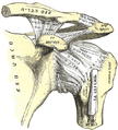
Gray1194 (german, colored).jpg 544 × 496; 209 KB
-
Gray1194 zh.png 550 × 482; 145 KB
-
Gray1194-ar.png 550 × 482; 168 KB
-
Gray1194.png 550 × 482; 122 KB
-
Gray1195 zh.png 531 × 500; 135 KB
-
Gray1195-ar.png 531 × 500; 155 KB
-
Gray200.png 600 × 188; 12 KB
-
Gray201 Arabic YM.png 700 × 259; 62 KB
-
Gray201.png 600 × 205; 16 KB
-
Gray326 heb.png 452 × 500; 86 KB
-
Gray326.png 452 × 500; 53 KB
-
Gray385 - Scalenus anterior.png 553 × 593; 234 KB
-
Gray386.png 600 × 477; 44 KB
-
Gray507.png 725 × 800; 145 KB
-
Gray808 - PT.svg 444 × 375; 1.68 MB
-
Gray808.png 592 × 500; 126 KB
-
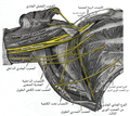
Gray809-ar.png 563 × 500; 160 KB
-
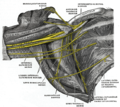
Gray809.png 563 × 500; 108 KB
-
Human clavicle.stl 5,120 × 2,880; 5.65 MB
-
KHRYSTYNA K. (8260332166).jpg 1,024 × 683; 373 KB
-
Left clavicle - close-up - animation.gif 450 × 450; 372 KB
-
Left clavicle - close-up - anterior view.png 4,500 × 4,500; 509 KB
-
Left clavicle - close-up - inferior view animation.gif 450 × 450; 546 KB
-
Left clavicle - close-up - inferior view.png 4,500 × 4,500; 765 KB
-
Left clavicle - close-up - lateral view.png 4,500 × 4,500; 773 KB
-
Left clavicle - close-up - medial view.png 4,500 × 4,500; 683 KB
-
Left clavicle - close-up - posterior view.png 4,500 × 4,500; 547 KB
-
Left clavicle - close-up - superior view animation.gif 450 × 450; 457 KB
-
Left clavicle - close-up - superior view.png 4,500 × 4,500; 735 KB
-
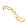
Left Clavicle.stl 5,120 × 2,880; 9.04 MB
-
Leonardo da vinci, studi anatomici 1509-1510.jpg 701 × 1,006; 114 KB
-
Male right shoulder and region towards neck, photographed when arm stretched out to side.jpg 2,700 × 1,600; 3.28 MB
-
Middle age Female Neck.png 7,378 × 4,924; 39.73 MB
-
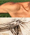
Photo of male right shoulder, combined with an anatomical drawing from Leonardo da Vinci.jpg 2,700 × 3,200; 3.51 MB
-
Processus conoideus der Klavikula 26M - CR ap - 001.jpg 804 × 777; 86 KB
-
Prominent conoid process of the clavicle 22jw - Roe ap - 001.jpg 1,146 × 1,128; 113 KB
-
Prominenter Processus conoideus der Clavicula 45M - CR ap - 001.jpg 1,964 × 1,407; 350 KB
-
Quain's elements of anatomy (1891) - Vol2 Part1- Fig 085.png 1,526 × 626; 572 KB
-
Quain's elements of anatomy (1891) - Vol2 Part1- Fig 086.png 1,548 × 594; 699 KB
-
Right clavicle - close-up - animation.gif 450 × 450; 370 KB
-
Right clavicle - close-up - anterior view.png 4,500 × 4,500; 506 KB
-
Right clavicle - close-up - inferior view animation.gif 450 × 450; 494 KB
-
Right clavicle - close-up - inferior view.png 4,500 × 4,500; 691 KB
-
Right clavicle - close-up - lateral view.png 4,500 × 4,500; 778 KB
-
Right clavicle - close-up - medial view.png 4,500 × 4,500; 682 KB
-
Right clavicle - close-up - posterior view.png 4,500 × 4,500; 531 KB
-
Right clavicle - close-up - superior view animation.gif 450 × 320; 441 KB
-
Right clavicle - close-up - superior view.png 4,500 × 4,500; 814 KB
-
Rotatorcuff.jpg 250 × 264; 10 KB
-
Scapula, clavicula - posterior (model).jpg 3,456 × 4,608; 4.17 MB
-
Shoulder and it's anatomy.webm 20 s, 480 × 360; 1.2 MB
-
Shoulder joint anatomy quiz.jpg 800 × 600; 111 KB
-
Shoulder joint-ar.svg 391 × 353; 339 KB
-
Shoulder joint-ca.svg 391 × 353; 356 KB
-
Shoulder joint-es.svg 391 × 353; 366 KB
-
Shoulder joint-pt.svg 391 × 353; 373 KB
-
Shoulder joint-zh.svg 391 × 353; 91 KB
-
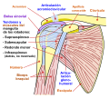
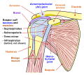
Shoulder joint.svg 391 × 353; 344 KB
-
Shoulderjoint.PNG 370 × 390; 26 KB
-
Slide8b.JPG 960 × 720; 120 KB
-
Sobo 1909 539.png 1,264 × 1,628; 7.86 MB
-
Sobo 1909 699.png 956 × 1,184; 3.24 MB
-
Spongiosa graft (6025357060).jpg 3,668 × 2,268; 1.24 MB
-
Starved Vietnamese man, 1966.JPEG 2,031 × 2,858; 1.33 MB
-
Structure of Adam's apple.png 531 × 500; 120 KB
-
Vue inférieure clavicule gauche.jpg 802 × 265; 126 KB
-
William Cheselden body.jpg 744 × 1,385; 315 KB
-
쇄골성장판.jpg 1,175 × 563; 33 KB
-
어깨 너비(쇄골 길이) 평균.jpg 770 × 390; 38 KB
_(14753370116).jpg/95px-A_treatise_on_dislocations_and_on_fractures_of_the_joints_(1824)_(14753370116).jpg)
_(14570815688).jpg/119px-American_quarterly_of_roentgenology_(1909)_(14570815688).jpg)







.jpg/120px-Collarbone_(PSF).jpg)


.jpg/120px-Disposo-T_--Clavicle_Splint_(NBY_428981).jpg)
_-_Fig_031.png/120px-Dixon%27s_Manual_of_human_osteology_(1912)_-_Fig_031.png)
_-_Fig._158.png/120px-Gerrish%27s_Text-book_of_Anatomy_(1902)_-_Fig._158.png)

.jpg/120px-Gray1194_(german%2C_colored).jpg)






.jpg/120px-KHRYSTYNA_K._(8260332166).jpg)


_-_Google_Art_Project.jpg/83px-Leonardo_da_Vinci_-_Superficial_anatomy_of_the_shoulder_and_neck_(recto)_-_Google_Art_Project.jpg)







.jpg/90px-Scapula%2C_clavicula_-_posterior_(model).jpg)






.jpg/120px-Spongiosa_graft_(6025357060).jpg)



_평균.jpg/120px-어깨_너비(쇄골_길이)_평균.jpg)